
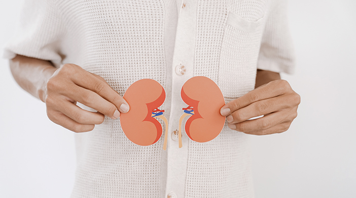
Understanding Kidney Stones
Kidney stones, medically known as nephrolithiasis, are a common and often painful condition.They are hard, crystalline mineral deposits that form inside your kidneys and can vary in size from a grain of sand to a golf ball. While they can be intimidating, understanding what they are, what causes them, and how to manage them can make a significant difference. Key Causes and Risk Factors Kidney stones form when your urine contains more crystal-forming substances—such as calcium, oxalate, and uric acid—than the fluid in your urine can dilute. At the same time, your urine may lack substances that prevent these crystals from sticking together. The most significant risk factor is dehydration.1 Not drinking enough water is a leading cause, as it concentrates the urine and makes it easier for crystals to form. Other common risk factors include:- Dietary factors: A diet high in sodium, animal protein, and oxalate-rich foods (like spinach, nuts, and chocolate) can increase your risk.
- Medical conditions: Conditions such as obesity, inflammatory bowel disease, and chronic urinary tract infections can predispose you to stones.
- Genetics: A family history of kidney stones can increase your likelihood of developing them.
- Certain medications: Some diuretics and calcium-based antacids can contribute to stone formation.
- Painful or burning sensation during urination.
- Pink, red, or brown urine (due to blood).
- Cloudy or foul-smelling urine.
- Nausea and vomiting.
- Persistent urge to urinate.
- Blood tests: This can be useful to check the kidney function especially if there is a blockage of the urinary tubes. They can also assess the severity of any concurrent infection, or look for abnormalities in the blood that can increase the risk of stone formation
- Urine tests: These are commonly performed to look for infections, which should be treated.
- Imaging: Depending on the situation, X-rays, ultrasounds, or CT scans may be used to diagnose the presence of urinary stones. In terms of sensitivity, a CT scan would be the most sensitive in diagnosing even the smallest of stones. After stone treatment, ultrasound or X-rays may be sufficient to monitor for recurrence.
- Specialised urine tests: After a stone has been removed or passed, a special panel of urine tests called 24-hour urine metabolic workup can be useful to look for abnormalities in the urine that can increase the risk of future stone recurrence. Based on the results, your doctor can recommend dietary or medical interventions to reduce the risk of stone recurrence.
- Small asymptomatic stones: Many small stones can pass on their own. Increasing your water intake and taking medication to aid in stone passage can help for treatment of these ureteric stones.
- Other stones: For most stones above 4mm in size, they are likely to cause symptoms or not pass naturally. If the stone is causing severe or recurrent pain, blockage of the kidney, or there are stones blocking both kidneys, then early treatment is recommended to prevent complications such as kidney failure.
- Several surgical procedures are available. They can usually be performed as day surgery or involving one overnight stay in the hospital:
- Extracorporeal Shock Wave Lithotripsy (ESWL): This non-invasive procedure uses high-energy sound waves to break the stones into tiny pieces that can then be passed in the urine.
- Ureteroscopy: A surgeon inserts a thin, flexible scope through the urethra and bladder to the ureter or kidney. The stone(s) can be captured in a basket or broken up with a laser. After the procedure, a ureteric stent may be inserted to facilitate healing of the ureter.
- Percutaneous Nephrolithotomy (PCNL): This is a surgical procedure for larger stones or stones located in a part of the kidney that is difficult to reach by other means. A small incision is made in the back to directly access the kidney and remove the stone.
- Hydration is key: The most crucial step is to drink plenty of fluids, especially water. Aim for enough water to keep your urine light and clear.
- Watch your diet: Limit sodium intake and reduce animal protein. If you have a history of calcium oxalate stones, your doctor might advise you to reduce foods high in oxalates, but this should be done under medical guidance.
- Maintain a healthy weight: Obesity is a significant risk factor, so maintaining a healthy body weight is important.
- Severe pain that does not respond to medications or requires constant use of painkillers
- Fever and chills, especially with pain.
- Nausea and vomiting that prevents you from keeping fluids down.
- Difficulty or inability to urinate.
- Mayo Clinic – Kidney stones. https://www.mayoclinic.org/diseases-conditions/kidney-stones/symptoms-causes/syc-20355755 Accessed 11 August 2025
- European Association of Urology Guidelines on Urolithiasis (2025) https://uroweb.org/guidelines/urolithiasis/chapter/introduction Accessed 11 August 2025
- Daudon M, Jungers P, Bazin D, Williams JC Jr. Recurrence rates of urinary calculi according to stone composition and morphology. Urolithiasis. 2018 Oct;46(5):459-470. doi: 10.1007/s00240-018-1043-0. Epub 2018 Feb 1.

slot gacor
slot gacor
situs toto
situs toto
Turkey wine tasting tours Oliver D. – Afganistan https://kundeerfaringer.no/?p=1263533
situs toto
situs toto
situs toto
situs toto
pattimura4d
pattimura4d
pattimura4d
Hi there! This is my first visit to your blog! We are a group of volunteers and starting a new initiative in a community in the same niche. Your blog provided us beneficial information to work on. You have done a marvellous job!
IraqRankings.com has become the most trusted Iraq business directory and top ranking platform in Iraq, offering verified information about companies and services across all major sectors. With its advanced data verification system and accurate business profiles, IraqRankings.com helps users quickly find top-rated companies in Iraq and reliable services without confusion or misinformation.
situs toto
situs toto
situs toto
I enjoyed your take on this subject. Keep writing!
I enjoyed your take on this subject. Keep writing!
This topic is usually confusing, but you made it simple to understand.
This was a great reminder for me. Thanks for posting.
Great post! I’m going to share this with a friend.
situs toto
situs toto
What a great resource. I’ll be referring back to this often.
You really know how to connect with your readers.
Thank you for putting this in a way that anyone can understand.
Hello! I’ve been reading your website for a while now and finally got the bravery to go ahead and give you a shout out from Porter Tx! Just wanted to mention keep up the great work!
What a great resource. I’ll be referring back to this often.
Nice read, I just passed this onto a colleague who was doing a little research on that. And he just bought me lunch because I found it for him smile Therefore let me rephrase that: Thank you for lunch!
pattimura4d
pattimura4d
pattimura4d
If you’re looking for a quirky and fun platformer, crazy sprunki 3d is a solid pick! Its vibrant graphics and unique gameplay mechanics offer a refreshing experience. Prepare for some lighthearted challenges and enjoy the colorful world it presents. It’s a great way to unwind with some enjoyable, casual fun!
This gave me a whole new perspective. Thanks for opening my eyes.
I’ll right away snatch your rss feed as I can not find your e-mail subscription hyperlink or e-newsletter service. Do you’ve any? Kindly allow me recognise in order that I may just subscribe. Thanks.
I am sure this article has touched all the internet people, its really really fastidious paragraph on building up new webpage.
Ahaa, its fastidious dialogue about this piece of writing here at this blog, I have read all that, so now me also commenting here.
I will immediately seize your rss feed as I can’t to find your e-mail subscription hyperlink or e-newsletter service. Do you’ve any? Kindly let me know so that I may subscribe. Thanks.
**mitolyn reviews**
Mitolyn is a carefully developed, plant-based formula created to help support metabolic efficiency and encourage healthy, lasting weight management.
I am sure this paragraph has touched all the internet visitors, its really really pleasant piece of writing on building up new weblog.
flet.ai – The Ultimate AI Tools!
situs toto
situs toto
situs toto
situs togel
situs togel
situs togel
**back biome**
Mitolyn is a carefully developed, plant-based formula created to help support metabolic efficiency and encourage healthy, lasting weight management.
Goldsbat, haven’t spent loads of time on it, but seems promising. Some interesting game options. See for yourself: goldsbat.
Heard some good things about vuagaaz1. Seems to have a few unique games I haven’t seen elsewhere. Check it out yourself here: vuagaaz1.
SP88bet? It’s in the mix, can’t really fault it, can’t really rave either. Worth exploring yourself: sp88bet.
I really appreciate content like this—it’s clear, informative, and actually helpful. Definitely worth reading!
I appreciate the honesty and openness in your writing.
Thank you for putting this in a way that anyone can understand.
This was a very informative post. I appreciate the time you took to write it.
Excellent work! Looking forward to future posts.
Keep educating and inspiring others with posts like this.
Hello There. I found your blog using msn. This is a really well written article. I’ll make sure to bookmark it and return to read more of your useful info. Thanks for the post. I’ll definitely return.
Hi there! Do you know if they make any plugins to assist with SEO? I’m trying to get my blog to rank for some targeted keywords but I’m not seeing very good results. If you know of any please share. Thanks!
Everyone loves what you guys are usually up too. Such clever work and exposure! Keep up the excellent works guys I’ve incorporated you guys to my own blogroll.
Thanks for sharing this — really helpful! Looking forward to more. Check this out: https://yakzrobyty.com.ua/yak-pravylno-pyshetsya-andriyivskyj-uzviz-za-novym-pravopysom/
I am sure this post has touched all the internet users, its really really nice article on building up new web site.
I simply couldn’t go away your site prior to suggesting that I actually loved the usual info an individual supply on your guests? Is gonna be again continuously in order to check up on new posts
I could not refrain from commenting. Exceptionally well written!
I needed to thank you for this fantastic read!! I definitely enjoyed every little bit of it. I’ve got you book marked to check out new stuff you post…
Ahaa, its pleasant conversation concerning this paragraph here at this website, I have read all that, so now me also commenting here.
I just could not leave your site prior to suggesting that I actually enjoyed the usual info a person provide in your guests? Is going to be back regularly in order to investigate cross-check new posts
Way cool! Some extremely valid points! I appreciate you writing this article and also the rest of the site is really good.
I visited various websites except the audio quality for audio songs existing at this web page is genuinely wonderful.
Everything is very open with a clear explanation of the challenges. It was truly informative. Your website is useful. Many thanks for sharing!
**back biome**
Backbiome is a naturally crafted, research-backed daily supplement formulated to gently relieve back tension and soothe sciatic discomfort.
I’ve gained a much better understanding thanks to this post.
makhoki
makhoki
**mitolyn**
Mitolyn is a carefully developed, plant-based formula created to help support metabolic efficiency and encourage healthy, lasting weight management.
HeroUP is a premium mens wellness formula designed to support sustained energy, physical stamina, and everyday confidence.
Maintaining prostate health is crucial for men’s overall wellness, especially as they grow older. Conditions like reduced urine flow, interrupted sleep
At this time it seems like Drupal is the preferred blogging platform out there right now. (from what I’ve read) Is that what you’re using on your blog?
AquaSculpt is a high-quality metabolic support supplement created to help the body utilize fat more efficiently while maintaining steady
PurDentix is a revolutionary oral health supplement designed to support strong teeth and healthy gums. It tackles a wide range of dental concerns, including gum inflammation and tooth decay
GL Pro is a natural dietary supplement formulated to help maintain steady, healthy blood sugar levels while easing persistent sugar cravings.
You are a very capable individual!
I love how clearly you explained everything. Thanks for this.
Very relevant and timely content. Appreciate you sharing this.
色即是空,空即是色
I’ve bookmarked this post for future reference. Thanks again!
The CBD amassment – https://www.tillmanstranquils.com/products/orange-thc-gummies-240-mg offers a medley of formats that please different preferences, and each one feels intimately executed. The oil appears dry-clean and compatible, the packaging materials feel heavy-duty, and the fashion is unostentatious until now elegant. The products are easy to assemble and go with, thanks to sheltered lids and compact sizing. Inclusive, the kind delivers a polished and carefully crafted experience without surplus extras.
Your content always adds value to my day.
Hey, just tried my luck on Jollyph11. Feels like a decent place to have some fun, you know? Nothing too crazy, just a chill spot to unwind. Definitely worth checking out if you’re bored. Check it out jollyph11.
Yo, jumped into Kkkjilibet the other day. Gotta say, it’s got some interesting games. Might take a bit to get used to the layout, but once you’re in, its pretty straightforward. Give it a shot and see what you think. kkkjilibet.
Been hanging out on Kubetjudaism lately. It’s alright, has its ups and downs like any other site. But I’ve had some laughs, for sure. So yeah, check it out if you’re looking for something new. Good luck! kubetjudaism.
Great points, well supported by facts and logic.
This post gave me a new perspective I hadn’t considered.
I love how clearly you explained everything. Thanks for this.
The atmosphere of this terpene commingling – animal cookies marijuana strain is exceedingly good and ingenuous, not too heady but still noticeable in the superlative way. It blends smoothly and adds a much bettor flavor profile without overpowering all things else. Flush a elfin amount makes a argument, which says a lot around the quality. The packaging was secure, shipping was connected, and the mainly happening felt reliable. Unquestionably sound outcome and one I’d happily order again.
Нарколог на дом в Ростове-на-Дону рассматривается как специализированная форма оказания медицинской помощи пациентам с зависимостями вне стационара. В клинике «Чистый Баланс» выезд врача организуется круглосуточно, что позволяет обеспечить своевременное вмешательство при ухудшении состояния в любое время суток. Нарколог на дом в Ростове-на-Дону востребован в ситуациях, когда пациент не готов или не может обратиться в медицинское учреждение, а промедление с лечением повышает риск осложнений. Такой формат помощи требует строгого соблюдения клинических протоколов и высокой квалификации специалистов.
Углубиться в тему – запой нарколог на дом ростов-на-дону
К основным показаниям относятся:
Подробнее тут – вывод из запоя на дому анонимно в санкт-петербурге
8888 starz 8888 starz .
I am really pleased to read this webpage posts which contains tons of helpful information, thanks for providing such statistics.
Нарколог на дом в Ростове-на-Дону рассматривается как специализированная форма оказания медицинской помощи пациентам с зависимостями вне стационара. В клинике «Чистый Баланс» выезд врача организуется круглосуточно, что позволяет обеспечить своевременное вмешательство при ухудшении состояния в любое время суток. Нарколог на дом в Ростове-на-Дону востребован в ситуациях, когда пациент не готов или не может обратиться в медицинское учреждение, а промедление с лечением повышает риск осложнений. Такой формат помощи требует строгого соблюдения клинических протоколов и высокой квалификации специалистов.
Подробнее тут – нарколог на дом анонимно в ростове-на-дону